Tweet
The last rickettsial disease I will cover at Infection Landscapes is Rocky Mountain spotted fever. This is not a collection of diseases as was the case with typhus and ehrlichiosis. Rather, it is a single and quite severe condition. Indeed, it is the most severe of all the rickettsial diseases.
Rocky Mountain spotted fever (RMSF) is caused by Rickettsia rickettsii, and is vectored by yet another tick. We will look at the pathogen and vector in turn before coming back to the clinical manifestations and the epidemiology of the disease.
The Pathogen: Rickettsia rickettsii is an obligate intracellular parasitic organism, as are all other Rickettsia and Rickettsiales bacteria:
This bacterium's target are endothelial cells, which it invades by a rather remarkable mechanism. The organism first adheres to the surface of the host endothelial cell and subsequently induces structural changes within, which causes the cell to phagocytize the Rickettsia organism. Since endothelial cells are not phagocytes, this bacterium has evolved a mechanism of entry into host cells by coercing them to perform functions they are not evolved to perform. This invasion of, and subsequent proliferation in, endothelial cells results in vascular damage and can lead to widespread organ failure.
The Vector: This rickettsial disease is again vectored by ticks. Dermacentor variabilis is the primary vector in the eastern and southern US, while D. andersoni is more common in the northwest:
Amblyomma cajennense is the primary vector in Central and South America:
To see how these ticks (Dermacentor in particular) compare to the other species we have covered at Infection Landscapes, here is the familiar scaled image:
Dermacentor is somewhat larger than the Ixodes species, but is similar in size to the Amblyomma species, though the Amblyomma are slightly more stout in appearance during their nymphal and adult life cycle stages. A. americanum and A. cajennense are similar in size, even though their dorsal markings can be quite different.
Here are the distributions of the three important tick vectors for RMSF:
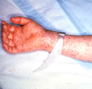
The Disease: This is the definitive rickettsial disease. It is characterized by the sudden onset of fever, chills, severe headache and myalgia, reddening of the eyes, and often debilitating malaise. This acute illness typically lasts 2 to 3 weeks when untreated. During the first week of illness a maculopapular rash often presents on the extremities, usually sometime between day 3 and 5. This rash can quickly spread to the palmar and plantar surfaces and subsequently extends to the trunk:
You will recall this is the opposite pattern to the rash observed in the typhus forms we discussed, which begins on the trunk and moves out to the extremities. In 40% to 60% of adults, or 90% of kids, the rash can become petachial exanthematous toward the end of the first week. Because R. rickettsii invades and damages endothelial cells, widespread organ damage involving the gut, lung, kidneys and the central nervous system can ensue. Thrombocytopenia and elevated liver enzymes can be important findings preceding these more extended systemic sequelae. The case-fatality ranges between 20% and 80% in untreated individuals, with older individuals being at significantly higher risk. With quick diagnosis and treatment the case-fatality is reduced to 3%-5%.
Although distributed throughout the Americas, the greatest number of cases of RMSF occur in the US. In addition, RMSF is the most commonly reported rickettsial disease in the US. Here is a map from the Centers for Disease Control and Prevention (CDC) showing the incidence of RMSF by state in 2008:
Notice the greatest incidence occurs in the south-central and southeastern seaboard regions of the country, despite the "Rocky Mountain" in the common name.
This CDC graph below, shows that rather than improving, RMSF has actually been increasing in incidence over the last two decades:
Given the similar incidence trends with other tick-borne infections, such as Lyme disease, we may do well to consider that more encounters between humans and ticks follow as urban development encroaches on natural habitat.
This concludes the short series on the rickettsial diseases. The series is by no means exhaustive, but I think it has served as a good introduction. Next week I will also be concluding the extended series on arthropod-borne infections, which began almost 6 months ago. It has been a fun and exciting series, and it will be concluded next week with yellow fever.
Following this, we will focus on two important airborne infections: measles and tuberculosis.
The last rickettsial disease I will cover at Infection Landscapes is Rocky Mountain spotted fever. This is not a collection of diseases as was the case with typhus and ehrlichiosis. Rather, it is a single and quite severe condition. Indeed, it is the most severe of all the rickettsial diseases.
Rocky Mountain spotted fever (RMSF) is caused by Rickettsia rickettsii, and is vectored by yet another tick. We will look at the pathogen and vector in turn before coming back to the clinical manifestations and the epidemiology of the disease.
The Pathogen: Rickettsia rickettsii is an obligate intracellular parasitic organism, as are all other Rickettsia and Rickettsiales bacteria:
Rickettsia rickettsii
This bacterium's target are endothelial cells, which it invades by a rather remarkable mechanism. The organism first adheres to the surface of the host endothelial cell and subsequently induces structural changes within, which causes the cell to phagocytize the Rickettsia organism. Since endothelial cells are not phagocytes, this bacterium has evolved a mechanism of entry into host cells by coercing them to perform functions they are not evolved to perform. This invasion of, and subsequent proliferation in, endothelial cells results in vascular damage and can lead to widespread organ failure.
The Vector: This rickettsial disease is again vectored by ticks. Dermacentor variabilis is the primary vector in the eastern and southern US, while D. andersoni is more common in the northwest:
Dermacentor variabilis
Amblyomma cajennense is the primary vector in Central and South America:
Amblyomma cajennense
To see how these ticks (Dermacentor in particular) compare to the other species we have covered at Infection Landscapes, here is the familiar scaled image:
Dermacentor is somewhat larger than the Ixodes species, but is similar in size to the Amblyomma species, though the Amblyomma are slightly more stout in appearance during their nymphal and adult life cycle stages. A. americanum and A. cajennense are similar in size, even though their dorsal markings can be quite different.
Here are the distributions of the three important tick vectors for RMSF:
Dermacentor variabilis distribution
Dermacentor andersoni distribution
Amblyomma cajennense distribution
The life cycles of the Dermacentor species and A. cajennense are quite similar to Ixodes and other Amblyomma species, in that they are 3 host ectoparasites, requiring a blood meal during the larval, nymphal, and adult life stages, which typically take approximately 2 years to complete. Rather than review the general life cycle for a third time, I will simply refer the reader to the Lyme disease or ehrlichiosis descriptions.
Importantly, tick hosts are not necessary for the maintenance of R. rickettsii in nature, as this pathogen is transmitted transovarially between ticks (you'll recall we saw transovarial maintenance before with mite-borne scrub typhus). Therefore, the ticks are the primary reservoir for this rickettsial disease. Nevertheless, transstadial transmission of this pathogen from ticks to rodents and dogs is also significant. This creates additional natural reservoirs of these mammals that, while not necessary for maintaining R. rickettsii in nature, do amplify the pathogen's transmission capacity. Humans, as is the case with most tick-borne infections, are dead-end hosts. Therefore, once infected, humans are not capable of transmitting the pathogen to non-infected ticks.
You will recall this is the opposite pattern to the rash observed in the typhus forms we discussed, which begins on the trunk and moves out to the extremities. In 40% to 60% of adults, or 90% of kids, the rash can become petachial exanthematous toward the end of the first week. Because R. rickettsii invades and damages endothelial cells, widespread organ damage involving the gut, lung, kidneys and the central nervous system can ensue. Thrombocytopenia and elevated liver enzymes can be important findings preceding these more extended systemic sequelae. The case-fatality ranges between 20% and 80% in untreated individuals, with older individuals being at significantly higher risk. With quick diagnosis and treatment the case-fatality is reduced to 3%-5%.
Although distributed throughout the Americas, the greatest number of cases of RMSF occur in the US. In addition, RMSF is the most commonly reported rickettsial disease in the US. Here is a map from the Centers for Disease Control and Prevention (CDC) showing the incidence of RMSF by state in 2008:
Notice the greatest incidence occurs in the south-central and southeastern seaboard regions of the country, despite the "Rocky Mountain" in the common name.
This CDC graph below, shows that rather than improving, RMSF has actually been increasing in incidence over the last two decades:
Given the similar incidence trends with other tick-borne infections, such as Lyme disease, we may do well to consider that more encounters between humans and ticks follow as urban development encroaches on natural habitat.
Control and prevention of RMSF is, of course, similar in approach to that outlined for both Lyme disease and ehrlichiosis. Still, it is worth restating here. The focus must be on the human point of contact with ticks. Attempts to intervene at the level of any of the other organisms (i.e. tick hosts) will, almost certainly, meet with failure because of the transovarial transmission of the pathogen between ticks. In addition, and as with other tick-borne infections, attempts to control favored host species of the relevant tick (whether Dermacentor or Amblyomma ticks) are extremely difficult because the secondary rodent and dog reservoirs are ubiquitous, highly adaptive, and can exploit a wide range of habitat. Moreover, because dogs can act as reservoirs, they have the capacity to bring the pathogen and its primary reservoir and vector (i.e. the tick) directly into the human domestic environment. Thus elimination of the bacteria is highly unlikely. Therefore, control and prevention is best aimed at human points of contact. Use of long-sleeved shirts and long pants are very effective control measures as these eliminate tick access to human skin. However, this approach may not be realistic for those that live, or work outdoors in endemic areas during the summer months. As such, individuals who do spend time outside during the summer months and are at risk of exposure to ticks, should practice regular body tick checks.
Here is a nice video on the proper way to remove ticks from the skin:
This concludes the short series on the rickettsial diseases. The series is by no means exhaustive, but I think it has served as a good introduction. Next week I will also be concluding the extended series on arthropod-borne infections, which began almost 6 months ago. It has been a fun and exciting series, and it will be concluded next week with yellow fever.
Following this, we will focus on two important airborne infections: measles and tuberculosis.


