
Tweet
This week at Infection Landscapes I am going to cover another nematode infection: strongyloidiasis. This infection is not nearly as prevalent as the three major soil-transmitted helminths, which, together, constitute probably the greatest global burden of disease attributable to the neglected tropical diseases. Nevertheless, strongyloidiasis currently infects up to 100 million people in the world, and can have very serious consequences among immunocompromised individuals. As such, this is both a substantive and serious disease for many people in the world, especially those in the developing world who are co-infected with the human immunodeficiency virus (HIV).
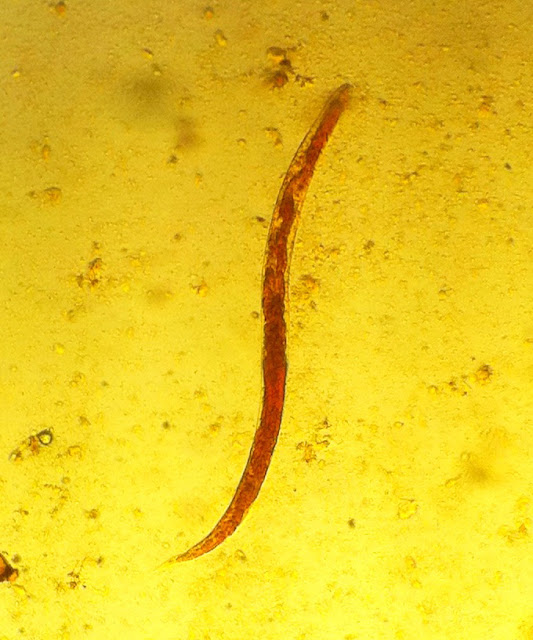
The Worm. Strongyloidiasis is caused by the nematode Strongyloides stercoralis:
This worm has one of the most complex, and fascinating, life cycles of all the nematodes we will consider. What makes this helminth particularly interesting is its ability to complete its life cycle as either a parasitic worm, or a free living worm in the soil. Let's take a close look at the stages of this worm's life cycle so we can fully appreciate its evolutionary adaptation.
There are two key features to this worm's life cycle that allow it to organize itself into two parallel developmental regimes such that it can either 1) live entirely outside, and without need of, the host, or 2) live entirely within the host without need of secondary or intermediate hosts. The former describes the free-living non-parasitic form of the worm, while the latter describes the autoinfective helminth. Moreover, the two forms do not define mutually exclusive life cycles. Rather, autoinfection can certainly lead to the introduction of free living organisms into the environment, and free-living organisms can produce infective larvae that are capable of infecting new susceptible hosts. Here is a nice graphic developed by the Centers for Disease Control and Prevention (CDC) that depicts the complex life cycle of S. stercoralis:

As a starting point in this life cycle let's examine the point of human infection. The infectious form of S. stercoralis is the filariform larva. These larvae initiate infection by penetrating the skin of the host, in much the same way that we saw hookworm initiates infection. These larvae migrate through the subcutaneous tissue to reach the capillary beds where they gain access to the circulation. By passive transport through the circulation they reach the capillary beds in contact with the alveoli in the lungs and migrate into these air sacs. From this position deep in the lungs the larvae migrate up the bronchioles, bronchii, and trachea, over the throat and are then swallowed. The larvae pass through the stomach and, upon reaching the small intestine, begin to mature into adults, where they will remain for the rest of the their life course as helminths woven into the villi of the epithelium. The adult females oviposit in the mucous membrane of the intestinal epithelium. Subsequently, after these eggs hatch, the 1st stage larvae emerge in the lumen of the small intestine and migrate to the large intestine. In the large intestine, the 1st stage larvae molt and transform into the rhabditiform larvae. This is a critical stage, as these rhabditiform larvae can follow one of three courses in the continued development of S stercoralis. They can 1) develop into filariform larvae, penetrate the gut wall, gain the circulation, remove to the alveoli, and, via migration up the respiratory tract, over the throat, and down the alimentary tract, regain the small intestine to begin a new infection in the adult stage. This intra-host infection cycle is known as autoinfection and can continue for many years through many generations of the worm; or 2) pass out of the host as rhabditiform larvae in the stool and develop into filariform larvae in the soil. These filariform larvae can then infect new hosts by penetrating the skin as described above; or 3) pass out of the host as rhabditiform larvae in the stool and develop into the next stage, again in the soil, but this time developing into free-living adults. These adults are not parasitic and so will complete their life course in the external environment with no need of a host. The free-living female adults oviposition directly in the soil where the eggs hatch and a new generation of rhabditiform larvae emerge. These rabditiform larvae can subsequently follow one of two courses to complete their life cycle. They can either develop into adults and continue as free living worms in the soil, or they can develop into filariform larvae and infect new hosts. The former, free-living, life cycle can continue indefinitely, producing subsequent cycles of free-living non-parasitic organisms. Whereas, a proliferation of filariform larvae are typically a response to unfavorable environmental cues such as a change in soil composition, moisture, or temperature, or a reduction in available food (i.e. bacteria) in the soil. However, with each subsequent generation of free-living worms, some of the rhabditiform larvae that emerge from the eggs will develop into filariform larvae so the potential for helminth development if the infectious larvae are able to contact a susceptible human host is always present even in the free-living community of S. stercoralis. Here again is the CDC graphic to help visualize this life cycle:
S. stercoralis is remarkable in that it has developed an opportunistic dual life course, such that it has the ability to exploit two distinct ecologic niches, engaging either as one or the other becomes available to the worm. Moreover, this organism maintains a bridge between these two disparate ecologic niches by way of the dual development potential of the free-living rhabditiform larvae. This is truly an extraordinarily adapted worm and a marvel of evolutionary biology. Unfortunately, S. stercoralis is capable of causing severe disease, so appreciation of the biology is quite tempered by the sobering morbidity for which this worm is responsible.
The Disease. Strongyloidiasis is comprised of a spectrum of disease. Many infections with S. stercoralis are asymptomatic. Because of the expansive migration of the worm throughout the human host, symptoms can present at multiple sites.
Common clinical disease includes 1) diarrhea, typically without dysentery, which results from the infection with the adult worms in the small intestine, 2) pulmonary symptoms in conjunction with the hypereosinophilia in the lungs, which results from the filariform migration into the alveoli and up the
respiratory tract, 3) hive-like rashes on the skin, which are a hypersensitivity reaction following the subcutaneous migration of the filariform larvae:

Strongyloidiasis can be either transient or chronic. Due to the autoinfection cycle described above, chronic infections do not necessarily require repeated points of contact with infectious filariform larvae in the environment. In other words, they do not require ongoing transmission. Nevertheless, the majority of chronic infections, more so than transient infections, are asymptomatic.
The S. stercoralis infections described thus far are, more or less, uncomplicated. Uncomplicated strongyloidiasis constitutes the large majority of the morbidity associated with this helminth. Whether symptomatic or asymptomatic, transient or chronic, most of these infections will not produce a severe life-threatening disease. However, complicated strongyloidiasis is a far more serious entity that can present in those with chronic infection.
Complicated, or disseminated, strongyloidiasis occurs when an individual with a chronic infection becomes immunocompromised. The suppressed immune response slackens the host's control over the infection and results in a hyperinfection, i.e. a massive proliferation S. stercoralis larvae. As the increased numbers of filariform larvae penetrate the epithelium of the large intestine and enter the circulation they likely carry some gut bacteria with them. This allows for the natural gut flora to access the blood and results in a dangerous septicemia. These bacteria can locate in organ systems throughout the body resulting in additional localized infection (thus the disseminated descriptor), with central nervous system involvement fairly common. Complicated strongyloidiasis can thus manifest with severe intestinal involvement directly due to the massive proliferation of larvae, or with disseminated disease due to the indirect transport of gut bacteria in the circulation. Complicated strongyloidiasis constitutes a life-threatening emergency and is associated with a high mortality when it occurs, which can be many years following the initial infection.
Individuals infected with HIV are at high risk of complicated strongyloidiasis if they are co-infected with S. stercoralis. Because strongyloidiasis is most prevalent in developing countries, these also may represent areas with a high occurrence of HIV infection depending on the specific location. Severe malnutrition can also lead to the kind to immunocompromise that would put someone with chronic strongyloidiasis at risk for complicated disease.
In addition, transplant patients who undergo immunosuppressive therapy following the transplant will be in danger of complicated strongyloidiasis if they are infected with S. stercoralis.
The Epidemiology and the Landscape. The current global prevalence of strongyloidiasis is estimated between a few tens of millions of cases to up to 100 million cases. These are primarily concentrated in the tropical and subtropical regions of the world, but can also be transmitted in some temperarte regions as well.

The highest prevalences of infection today are found in South and Southeast Asia, and in parts of Central and South America. Strongyloidiasis is endemic in much of Africa but the prevalence is typically quite low throughout the continent.
As we saw with both hookworm and whipworm, the range of S. stercoralis is determined by important aspects of the physical landscape and because of this, as well as critical overlapping characteristics of the human social landscape, the occurrence of strongyloidiasis follows similar lines of geography. Soil and climate are two critical landscape features that influence the distribution of S. stercoralis. Because passaged rhabditiform larvae can develop into free-living adults, and maintain a free-living cycle indefinitely, the non-parasitic ecology is distinctly characterized by specific environmental requirements. Whether free living or parasitic, the rhabditiform larvae that pass out in the host require sandy, loamy soils in order to develop into either filariform larvae, which can then infect humans, or into free-living adults. In addition, the soils must be moist and the temperature must be warm. As such, the specific climatic conditions limit the range of the worms to the tropical and subtropical regions of the world that receive significant amounts of precipitation on an annual basis, while the pedological and edaphological constraints further define the microgeography of these worms. Notice below the global distribution of soil morphology in the map produced by the Natural Resources Conservation Service (NRCS) of the United States Department of Agriculture:

And this NRCS map below depicting the global distribution of soil moisture:


Wearing good shoes without holes while outside in endemic areas is another critical step in the prevention of new S. stercoralis infections. Unfortunately, footwear is often simply not available for those people who need it most, and as such this very simple transmission block cannot be utilized.

Also, changing agricultural practices that rely on human feces for fertilization of crops could dramatically help reduce the distribution of S. stercoralis in soils in many agricultural subsistence communities.

Unfortunately this, too, can be a difficult practice to disengage since human feces serves as a very rich fertilizer and, thus, can form a critical component to subsistence farming in many parts of the world where other fertilizers or farming technologies are cost prohibitive. And, of course, without an affordable substitute, refraining from human feces fertilization could very well lead to starvation.
Finally, the unique circumstances of immunosuppression that lead to severe disease require that strongyloidiasis is given special consideration under conditions of malnutrition and/or co-infection with HIV or other immunosuppressive infections such as measles. Because all of these are of greater prevalence in the developing world where strongyloidiasis can also be endemic, severe disease is much more likely to affect these areas, especially in children. As such, it would be good practice to screen, and treat if necessary, individuals who have immunocompromising conditions in areas where strongyloidiasis is endemic.
This week at Infection Landscapes I am going to cover another nematode infection: strongyloidiasis. This infection is not nearly as prevalent as the three major soil-transmitted helminths, which, together, constitute probably the greatest global burden of disease attributable to the neglected tropical diseases. Nevertheless, strongyloidiasis currently infects up to 100 million people in the world, and can have very serious consequences among immunocompromised individuals. As such, this is both a substantive and serious disease for many people in the world, especially those in the developing world who are co-infected with the human immunodeficiency virus (HIV).
The Worm. Strongyloidiasis is caused by the nematode Strongyloides stercoralis:

This worm has one of the most complex, and fascinating, life cycles of all the nematodes we will consider. What makes this helminth particularly interesting is its ability to complete its life cycle as either a parasitic worm, or a free living worm in the soil. Let's take a close look at the stages of this worm's life cycle so we can fully appreciate its evolutionary adaptation.
There are two key features to this worm's life cycle that allow it to organize itself into two parallel developmental regimes such that it can either 1) live entirely outside, and without need of, the host, or 2) live entirely within the host without need of secondary or intermediate hosts. The former describes the free-living non-parasitic form of the worm, while the latter describes the autoinfective helminth. Moreover, the two forms do not define mutually exclusive life cycles. Rather, autoinfection can certainly lead to the introduction of free living organisms into the environment, and free-living organisms can produce infective larvae that are capable of infecting new susceptible hosts. Here is a nice graphic developed by the Centers for Disease Control and Prevention (CDC) that depicts the complex life cycle of S. stercoralis:

As a starting point in this life cycle let's examine the point of human infection. The infectious form of S. stercoralis is the filariform larva. These larvae initiate infection by penetrating the skin of the host, in much the same way that we saw hookworm initiates infection. These larvae migrate through the subcutaneous tissue to reach the capillary beds where they gain access to the circulation. By passive transport through the circulation they reach the capillary beds in contact with the alveoli in the lungs and migrate into these air sacs. From this position deep in the lungs the larvae migrate up the bronchioles, bronchii, and trachea, over the throat and are then swallowed. The larvae pass through the stomach and, upon reaching the small intestine, begin to mature into adults, where they will remain for the rest of the their life course as helminths woven into the villi of the epithelium. The adult females oviposit in the mucous membrane of the intestinal epithelium. Subsequently, after these eggs hatch, the 1st stage larvae emerge in the lumen of the small intestine and migrate to the large intestine. In the large intestine, the 1st stage larvae molt and transform into the rhabditiform larvae. This is a critical stage, as these rhabditiform larvae can follow one of three courses in the continued development of S stercoralis. They can 1) develop into filariform larvae, penetrate the gut wall, gain the circulation, remove to the alveoli, and, via migration up the respiratory tract, over the throat, and down the alimentary tract, regain the small intestine to begin a new infection in the adult stage. This intra-host infection cycle is known as autoinfection and can continue for many years through many generations of the worm; or 2) pass out of the host as rhabditiform larvae in the stool and develop into filariform larvae in the soil. These filariform larvae can then infect new hosts by penetrating the skin as described above; or 3) pass out of the host as rhabditiform larvae in the stool and develop into the next stage, again in the soil, but this time developing into free-living adults. These adults are not parasitic and so will complete their life course in the external environment with no need of a host. The free-living female adults oviposition directly in the soil where the eggs hatch and a new generation of rhabditiform larvae emerge. These rabditiform larvae can subsequently follow one of two courses to complete their life cycle. They can either develop into adults and continue as free living worms in the soil, or they can develop into filariform larvae and infect new hosts. The former, free-living, life cycle can continue indefinitely, producing subsequent cycles of free-living non-parasitic organisms. Whereas, a proliferation of filariform larvae are typically a response to unfavorable environmental cues such as a change in soil composition, moisture, or temperature, or a reduction in available food (i.e. bacteria) in the soil. However, with each subsequent generation of free-living worms, some of the rhabditiform larvae that emerge from the eggs will develop into filariform larvae so the potential for helminth development if the infectious larvae are able to contact a susceptible human host is always present even in the free-living community of S. stercoralis. Here again is the CDC graphic to help visualize this life cycle:
S. stercoralis is remarkable in that it has developed an opportunistic dual life course, such that it has the ability to exploit two distinct ecologic niches, engaging either as one or the other becomes available to the worm. Moreover, this organism maintains a bridge between these two disparate ecologic niches by way of the dual development potential of the free-living rhabditiform larvae. This is truly an extraordinarily adapted worm and a marvel of evolutionary biology. Unfortunately, S. stercoralis is capable of causing severe disease, so appreciation of the biology is quite tempered by the sobering morbidity for which this worm is responsible.
The Disease. Strongyloidiasis is comprised of a spectrum of disease. Many infections with S. stercoralis are asymptomatic. Because of the expansive migration of the worm throughout the human host, symptoms can present at multiple sites.
Common clinical disease includes 1) diarrhea, typically without dysentery, which results from the infection with the adult worms in the small intestine, 2) pulmonary symptoms in conjunction with the hypereosinophilia in the lungs, which results from the filariform migration into the alveoli and up the
respiratory tract, 3) hive-like rashes on the skin, which are a hypersensitivity reaction following the subcutaneous migration of the filariform larvae:

(A) Strongyloides pneumonitis associated with hyperinfection in a kidney transplant recipient. (B) Migrating larvae in subcutaneous lymphatics (arrows). (C) Hatching eggs in human intestine. (Published in: Parasitic infections in transplant recipients. Rashad S Barsoum. Nature Clinical Practice Nephrology (2006) 2, 490-503 doi:10.1038/ncpneph0255)
Strongyloidiasis can be either transient or chronic. Due to the autoinfection cycle described above, chronic infections do not necessarily require repeated points of contact with infectious filariform larvae in the environment. In other words, they do not require ongoing transmission. Nevertheless, the majority of chronic infections, more so than transient infections, are asymptomatic.
The S. stercoralis infections described thus far are, more or less, uncomplicated. Uncomplicated strongyloidiasis constitutes the large majority of the morbidity associated with this helminth. Whether symptomatic or asymptomatic, transient or chronic, most of these infections will not produce a severe life-threatening disease. However, complicated strongyloidiasis is a far more serious entity that can present in those with chronic infection.
Complicated, or disseminated, strongyloidiasis occurs when an individual with a chronic infection becomes immunocompromised. The suppressed immune response slackens the host's control over the infection and results in a hyperinfection, i.e. a massive proliferation S. stercoralis larvae. As the increased numbers of filariform larvae penetrate the epithelium of the large intestine and enter the circulation they likely carry some gut bacteria with them. This allows for the natural gut flora to access the blood and results in a dangerous septicemia. These bacteria can locate in organ systems throughout the body resulting in additional localized infection (thus the disseminated descriptor), with central nervous system involvement fairly common. Complicated strongyloidiasis can thus manifest with severe intestinal involvement directly due to the massive proliferation of larvae, or with disseminated disease due to the indirect transport of gut bacteria in the circulation. Complicated strongyloidiasis constitutes a life-threatening emergency and is associated with a high mortality when it occurs, which can be many years following the initial infection.
Individuals infected with HIV are at high risk of complicated strongyloidiasis if they are co-infected with S. stercoralis. Because strongyloidiasis is most prevalent in developing countries, these also may represent areas with a high occurrence of HIV infection depending on the specific location. Severe malnutrition can also lead to the kind to immunocompromise that would put someone with chronic strongyloidiasis at risk for complicated disease.
In addition, transplant patients who undergo immunosuppressive therapy following the transplant will be in danger of complicated strongyloidiasis if they are infected with S. stercoralis.
The Epidemiology and the Landscape. The current global prevalence of strongyloidiasis is estimated between a few tens of millions of cases to up to 100 million cases. These are primarily concentrated in the tropical and subtropical regions of the world, but can also be transmitted in some temperarte regions as well.

The highest prevalences of infection today are found in South and Southeast Asia, and in parts of Central and South America. Strongyloidiasis is endemic in much of Africa but the prevalence is typically quite low throughout the continent.
As we saw with both hookworm and whipworm, the range of S. stercoralis is determined by important aspects of the physical landscape and because of this, as well as critical overlapping characteristics of the human social landscape, the occurrence of strongyloidiasis follows similar lines of geography. Soil and climate are two critical landscape features that influence the distribution of S. stercoralis. Because passaged rhabditiform larvae can develop into free-living adults, and maintain a free-living cycle indefinitely, the non-parasitic ecology is distinctly characterized by specific environmental requirements. Whether free living or parasitic, the rhabditiform larvae that pass out in the host require sandy, loamy soils in order to develop into either filariform larvae, which can then infect humans, or into free-living adults. In addition, the soils must be moist and the temperature must be warm. As such, the specific climatic conditions limit the range of the worms to the tropical and subtropical regions of the world that receive significant amounts of precipitation on an annual basis, while the pedological and edaphological constraints further define the microgeography of these worms. Notice below the global distribution of soil morphology in the map produced by the Natural Resources Conservation Service (NRCS) of the United States Department of Agriculture:

And this NRCS map below depicting the global distribution of soil moisture:


And, finally, the map below by the United Nations Food and Agriculture Organization depicts the global distribution of the annual mean temperature:

It is worth noting how closely the global distribution of S. stercoralis coincides with the global distributions of soil regimes, moisture, and temperature. Finally, the filariform larvae are able to survive in, and are quite motile in, water, which greatly increases their versatility and transmission potential in comparison with hookworm.
There are three important factors from the S. stercoralis life cycle that are critical to the landscape epidemiology of human transmission. First, the rhabditiform larvae pass out into the environment in the feces of the human host. Second, these larvae live in the soil and can either develop into free-living adults, OR develop into filariform larvae, which can infect humans. Third, infectious filariform larvae must make contact with the skin of a new human host. These three factors determine how the social landscape intersects with the physical landscape to enable transmission to humans.
Lack of sanitation infrastructure, and especially a means by which human waste can be removed from sites of human occupation, results in feces being distributed directly in the human environment or in proximal spaces. Also, poor water quality is important for transmission. Conditions of poverty that are associated with the lack of municipal resources for infrastructural development often coincide with a lack of personal resources for adequate waste disposal, adequate water, and adequate clothing. As such, repeated exposure to filariform larvae is common in the same human environments (both the home and places of work) in which rhabditiform larvae-laden human feces are deposited on a daily basis. This leads to an abundance of points of contact for transmission between S. stercoralis and human hosts in those intersecting landscapes of warm, moist, structurally rich soils and conditions of poverty. This intersection currently defines a geography that encompasses, almost exclusively, the developing world.
In many poor subsistence agricultural communities, farmers use human feces as a fertilizer to enhance the growth of their crops. This readily available fertilizer provides a cheap, yet very rich, source of critical nutrients to the soil, which can mean the difference between a crop yield that provides the farmer with a livelihood and a yield that does not. Unfortunately, in areas where strongyloidiasis is endemic, the use of human feces as fertilizer means a constant and widespread distribution of rhabditiform larvae throughout the farming community, and thus a steady source of new infections.
Control and Prevention. Control and prevention of strongyloidiasis begins by following the usual guidelines: improving sanitation and water infrastructure in resource poor areas. In most settings in the world where strongyloidiasis is a significant problem, improved infrastructure that can adequately remove human feces from the spaces of human occupation is a first priority in its prevention.
Where large-scale municipally-resourced sanitation infrastructure is not available, individual pit privies can be constructed for single homes, or clusters of homes. Here is a graphic that depicts the dimensions and structural components of such a privy:


Photo by Peter Byrne
Also, changing agricultural practices that rely on human feces for fertilization of crops could dramatically help reduce the distribution of S. stercoralis in soils in many agricultural subsistence communities.

Unfortunately this, too, can be a difficult practice to disengage since human feces serves as a very rich fertilizer and, thus, can form a critical component to subsistence farming in many parts of the world where other fertilizers or farming technologies are cost prohibitive. And, of course, without an affordable substitute, refraining from human feces fertilization could very well lead to starvation.
Finally, the unique circumstances of immunosuppression that lead to severe disease require that strongyloidiasis is given special consideration under conditions of malnutrition and/or co-infection with HIV or other immunosuppressive infections such as measles. Because all of these are of greater prevalence in the developing world where strongyloidiasis can also be endemic, severe disease is much more likely to affect these areas, especially in children. As such, it would be good practice to screen, and treat if necessary, individuals who have immunocompromising conditions in areas where strongyloidiasis is endemic.